What's new

China Science & Technology Forum

Physics - Focus: More Voltage from Bending Silicone Rubber
April 12, 2019• Physics 12, 42

The voltage generated by bending a flexible material is usually small, but a new trick can dramatically increase the effect.


X. Wen et al., Phys. Rev. Lett. (2019)
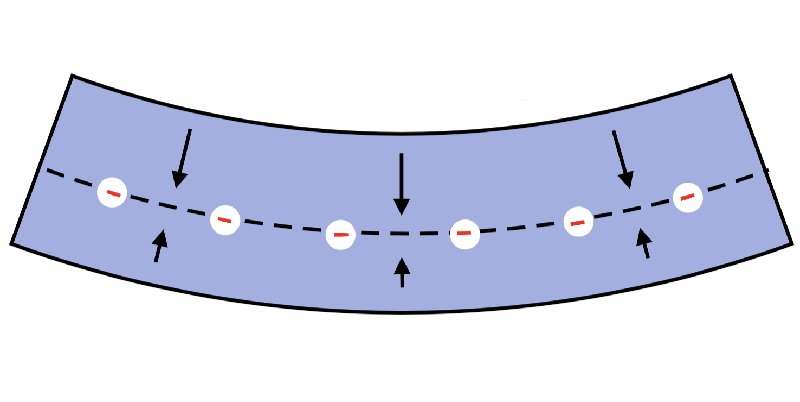
Battery power from flexing. Adding a charge layer to silicone rubber dramatically increases its ability to produce a voltage in response to being bent, which could lead to practical devices fairly soon. Bending the material squeezes the field lines in the upper half, increasing the electric field there (arrows), while spreading the field lines and reducing the field in the lower half.


Flexoelectric materials generate a voltage when bent, a property that could be useful in engineering delicate sensors or energy harvesting devices, such as clothes that would produce electricity when a person walks. Researchers have now shown that adding a layer of charge to the middle of a flexible polymer bar can boost the effect by 100 times. The team says that with further development, the effect could be used in real devices within five years.

Many ordinary materials such as crystals and polymers exhibit the flexoelectric effect. Bending the material causes each atomic layer to be stretched by a different amount, with the outermost layer stretched the most. This variation in stretch (a strain gradient) can create asymmetry in the positions of the material’s ions and can prevent the positive and negative charges from completely canceling, or, in other words, the material can become polarized. The polarization leads to a net electric field and thus a voltage.

Ceramics show the strongest effect, reflected in a high flexoelectric coefficient—a parameter indicating how much voltage is generated for a given bend. However, ceramics are brittle and can break from even small deformations. So researchers attempting to exploit the effect in practical devices have mostly focused on films of nanoscale thickness, which are easier to bend than thicker samples.

To produce the effect in macroscopic samples, one can use more flexible materials such as polymers, although their intrinsic flexoelectric effect is small. But now a team of researchers from Xi'an Jiaotong University in China has shown how to boost the effect in a polymer bar, merely by embedding a layer of permanent electric charge within the polymer.

Team leader Qian Deng and his colleagues experimented with a 10-cm-long bar of polydimethylsiloxane (PDMS, a type of silicone rubber) with width 15 mm and thickness 10 mm. The team embedded a thin layer of negatively charged polymer into the central plane along the length of the bar. This charge layer produced an electric field that created a voltage at the surfaces above and below the central plane.

The researchers then measured the flexoelectric coefficient for different amounts of embedded charge. In each measurement, with the bar horizontal, supported at each end, they applied a controlled downward force at the center, to deform the bar, and then measured the change in voltage registered at the surface.

The flexoelectric coefficient grew in direct proportion to the embedded charge. With charge sufficient to generate 5 kV on the polymer surface, the coefficient was 100 times larger than for PDMS without any embedded charge. The technique works, the researchers argue, because the parallel, vertical field lines emanating from the charge layer become splayed when the bar is bent. The lines spread out below the central plane and are compressed above it. This asymmetry leads to a strong polarization, measurable as a voltage across the bar, from bottom to top. The experimental results agreed closely with the team’s calculations.

“The basic idea is that the charge creates an initial electric field in the material, symmetric on the two sides,” Deng says. “Bending breaks this symmetry, so there's a net electric polarization across the film.”

“The effect the authors are seeing is substantial,” says nanoscience expert Gustau Catalán of the Catalan Institute of Nanoscience and Nanotechnology in Barcelona, Spain. He thinks the results could have broad implications. “This work has an appeal that goes beyond the flexoelectric community, and it could stimulate the search for similar effects in systems where, a priori, one would not have looked for flexoelectricity.”

Deng and colleagues believe the effect will find use in practical devices fairly soon. Although, according to team member Xin Wen, a key challenge will be learning how to prevent the loss of the embedded charge in the film, as it tends to slowly leak out.

As for commercial uses, Wen expects practical devices “in the next five years,” assuming input from a range of experts in the development process. He suggests that the effect will be useful in building devices such as sensors, energy harvesters, and actuators.


This research is published in Physical Review Letters.

–Mark Buchanan
Mark Buchanan is a freelance science writer who splits his time between Wales, UK, and Normandy, France.

Flexoelectret: An Electret with a Tunable Flexoelectriclike Response
Xin Wen, Dongfan Li, Kai Tan, Qian Deng, and Shengping Shen
Phys. Rev. Lett. 122, 148001 (2019)

Published April 12, 2019​
 
A new graphene foam stays squishy at the coldest temperatures
The superelastic material could be useful in space

BY MARIA TEMMING
2:00PM, APRIL 12, 2019

041119_mt_graphene_feat_rev.jpg
DEEP FREEZE Normal materials become brittle in deep cryogenic conditions, but a new graphene-based foam (shown in this scanning electron microscopy image) stays as flexible as ever.
K. ZHAO/SCIENCE ADVANCES 2019


A new graphene-based foam is the first material to remain soft and squishy even at deep cryogenic temperatures.

Most materials become stiff and brittle in extreme cold. But the new foam stays superelastic even when it’s subjected to the temperature of liquid helium: –269.15° Celsius. A material that remains pliable at such low temperatures could be used to build devices for use in space, researchers report online April 12 in Science Advances.

Inside this foam, oxygen atoms connect micrometer-sized patches of the superthin 2-D material graphene to create a meshlike structure (SN: 8/13/11, p. 26). The resulting material is flexible in deep cryogenic conditions because, even at such low temperatures, sheets of graphene are easily bendable and resistant to tearing, and the carbon-oxygen bonds that link these sheets together remain strong.

Yongsheng Chen, a materials scientist at Nankai University in Tianjin, China, and colleagues compressed samples of the material repeatedly at different temperatures. At –269.15° C, the foam behaved just as it did at room temperature, bouncing back to almost full size even after being compressed to one-tenth its original thickness. The material kept this resilience even when heated to about 1000° C and flattened hundreds of times.

Chen’s team suspects that different superthin materials, like 2-D semiconductors (SN Online: 2/13/18) or 2-D inorganic compounds (SN Online: 9/21/18) may create foams that might boast other unique properties.

Citations
K. Zhao et al. Super-elasticity of three-dimensionally cross-linked graphene materials all the way to deep cryogenic temperatures. Science Advances. Published online April 12, 2019. doi:10.1126/sciadv.aav2589.​


A new graphene foam stays squishy at the coldest temperatures | Science News
 
Engineered knee cartilage goes anisotropic
15 Apr 2019 Belle Dumé

Graphical abstract of the present study. Courtesy: D Jiang, Institute of Sports Medicine of Peking University Third Hospital

Researchers in China have succeeded in engineering knee cartilage that, for the first time, mimics the anisotropic characteristic of native tissue. The fabrication technique employed, which involves simultaneously applying biochemical and biomechanical stimuli to stem cells seeded onto a biomimetic scaffold, causes fibrochondrocytes in the bioconstruct to differentiate into layers containing two types of collagen. When transplanted in the knee joints of rabbits the material not only improves tensile strength in the knee after 24 weeks but also reduces joint cartilage degradation.

Although tissue engineering has come along in leaps and bounds over the past decade, most techniques still cannot faithfully reproduce the anisotropic nature of physiological systems that consist of heterogenous masses of connective tissue cells and an extracellular matrix (ECM).

The knee meniscus, which is an example of an anisotropic tissue, is a pad of cartilage that absorbs shocks and protects the knee from friction. It cannot fully heal after being damaged or torn, so patients suffering from such injuries would benefit from transplants of a biomimetic cartilage material.

Complex structure
The structure of the knee meniscus is complex however and is made up of outer and inner regions. In the outer region, fibroblast-like cells are contained with an ECM that is mainly made up of type I collagen, which makes the cartilage resistant to tensile loads. The inner region contains chondrocyte-like cells embedded within an ECM mainly made up of type II collagen and glycosaminoglycans, which make the tissue resistant to compression.

Until now, most tissue engineering techniques to reconstruct the knee meniscus were only able to produce homogenous issue. This tissue cannot withstand tensile or compressive stress and so degenerates over time.

Researchers led by Dong Jiang from the Institute of Sports Medicine of Peking University Third Hospital in China has now developed a technique in which they culture bone-marrow-derived mesenchymal stem cells (BMSCs) on a biomimetic scaffold. During the culture they apply two synergistic biochemical growth factors (cytokines) to the tissue and a loading system that simultaneously exerts both tensile and compressive stresses. The method induces the stem cells to differentiate into sperate layers of type I and type II collagen, thus mimicking the anisotropy in natural knee cartilage.

Jiang and colleagues say they have successfully tested their material on rabbits by transplanting it into the knee joints of the animals.

Long-term knee chondroprotection
“To our knowledge, ours is the first study to apply orchestrated biomechanical and biomechanical cues to make an anisotropic knee meniscus, as well as the first in vivo demonstration of an anisotropic-engineered meniscus for long-term knee chondroprotection,” says Jiang.

‘Qin Se He Mong’. Courtesy: D Jiang, Institute of Sports Medicine of Peking University Third Hospital

“Interestingly, our approach has an analogy in the Chinese idiom ‘Qin Se He Mong’, which describes the harmonious concerto produced by the two traditional musical instruments, Qin and Se,” he tells Physics World. “The note produced by the instruments could be represented by the biochemical and biomechanical stimuli respectively. Here, the synergy between both promotes the proliferation and differentiation of the BMSCs and reconstructs the anisotropic structures of the knee meniscus.”

Full details of the research are reported in Science Translational Medicine 10.1126/scitranslmed.aao0750.


Engineered knee cartilage goes anisotropic – Physics World
 
Ruthenium-based Catalyst Helps Water Electrolysis Rush
Apr 15, 2019

Hydrogen fuel, with the advantages of clean, renewable and of high fuel efficiency, is seen as the "ultimate fuel" and getting more attention around the world. Water electrolysis is an ideal way to produce hydrogen, but it requires active and stable catalysts which make this process more efficient and cheaper.

In a recent study published in Nature Catalysis as the cover story, Prof. WU Yuen's team from the University of Science and Technology of China (USTC) of Chinese Academy of Sciences successfully prepared a kind of Ruthenium (Ru) single atom alloy catalyst, which greatly accelerates the process of water eletrolysis with lower overpotential (220 mV).

By surface defect engineering to capture and stabilize single atoms, this single-atom catalyst is capable of delivering a 90 mV lower overpotential to reach a current density of 10 mA/cm2, and an order of magnitude with longer lifetime than that of commercial RuO2.

Researchers constructed a series of alloy-supported Ru1 in this study using different PtCu alloys through sequential acid etching and electrochemical leaching.

They also found a volcano relation between oxygen evolution reaction (OER) activity and the lattice constant of the PtCu alloys. Through density functional theory investigation, they revealed that the compressive strain of the Pt-skin shell engineers the electronic structure of the Ru1, allowing optimized binding of oxygen species and better resistance to over-oxidation and dissolution.

Compared with Iridium-based systems which have better dissolution resistance, Ru-based ones have more abundant reserves and have been evaluated to be a more active OER catalyst due to its lower overpotential.

This study makes hydrogen production through water electrolysis easier and more efficient, and allows people to see the great potential of hydrogen as an alternative new energy in the future.

Nevertheless, till now, the stability problem of catalyst hasn't been completely resolved, and the reaction system needs further improvement.

W020190415493175387289.jpg
Oxygen evolution reaction (OER) and oxygen reduction reaction (ORR) are regarded as two trophies in the field of efficient utilization of hydrogen energy. With Ru-based catalyst, acidic OER process is significantly accelerated. (Image by CUI Jie)

W020190415493175454864.jpg
In acidic electrolyte environment, water molecules was adsorbed on the active site Ru atom, and then electrolyzed. Purple, blue, red and white balls represents Ru, Pt, O and H atoms, respectively. (Image by WU Yuen)


Ruthenium-based Catalyst Helps Water Electrolysis Rush---Chinese Academy of Sciences

0ff4bdfd-350f-46e5-8667-19ada66a3d2b.png
 
Across China: Chinese doctors use 3D printing to make bone tissue
Source: Xinhua| 2019-04-17 01:19:41|Editor: Mu Xuequan

CHANGSHA, April 16 (Xinhua) -- Chinese surgeons in a central China hospital have succeeded in performing an implant surgery to replace bone tissue using 3D printing technology.

The 39-year-old patient surnamed Tan suffered from second phrase femoral head necrosis, said Hu Yihe, an orthopedic professor with Xiangya Hospital of Central South University in Hunan province.

"Walking was really painful, I could only lie in bed all day," Tan said.

According to doctors, the structure of Tan's bone trabecula -- the beam of the bone -- was damaged, and a new beam was needed.

"Hip joint replacement is the common practice, but the patient was at an early stage and wanted to keep his hip joint, so we decided to make a porous 3D printed tantalum scaffold which could support the damaged head of his femur and save his hip joint," Hu said.

Hu and his team built the computer 3D model based on Tan's imaging data and used 3D printing technology to customize the scaffold. They also made a 3D printed guide plate which assisted in finding the precise location during the surgery, shortening the operation time to less than 30 minutes.

The patient was able to stand the second day after the surgery and barely felt any pain.

After repeatedly placing his left foot on the right leg, and then vice-versa with ease, Tan was discharged from the hospital just three days later.

Hu and his team have worked with the clinical use of 3D printing for four years, and Tan's surgery on April 4 was the first application.

"The cause of femoral head necrosis and its radical treatment have so far remained unknown. For young patients at early stages, we hope to save their hip joints and help them lead a normal life by using new materials and methods such as 3D printing," Hu said.

3D printing has been embraced by doctors in many of China's major hospitals for surgeries and training, as the technology dramatically improves surgery precision.

In 2017, doctors in the Second Xiangya Hospital of Central South University in Hunan used 3D printing technology and performed two successful complex pediatric heart surgeries.

"3D printing gives patients more choices and has provided great new thoughts in clinical surgeries," Hu said.

12qno4s705214p768pn3nopn293p299p.jpg
 
18 Apr 2019 | 18:07 GMT
Better Ultrasound Imaging and Sonar Through Samarium
Samarium can nearly double the performance of piezoelectric crystals used in many sensors

By Charles Q. Choi

MzI3ODkyNg.jpeg
Photo: iStockphoto

Piezoelectric crystals are the key ingredient in many kinds of sensors that detect vibrations, and can be found in underwater sonars and medical ultrasound imaging systems. The crystals’ performance can be dramatically improved, according to a team of researchers at Xi'an Jiaotong University in China, by adding trace amounts of a rare-earth element.

Piezoelectric materials can convert mechanical oscillations to electrical signals and vice versa. Currently, the most advanced piezoelectric devices often use a perovskite oxide crystal known as PMN-PT, which outperforms other common piezoelectric materials by roughly a factor of three in terms of efficiency. However, despite much research, progress toward improving the performance of these crystals has been slow over the past two decades, researchers say.

Now scientists have discovered that introducing relatively minuscule amounts of samarium into PMN-PT—adding about one atom per thousand atoms of the parent crystals—could greatly enhance its performance. Regular PMN-PT crystals generate about 1,200 to 2,500 picocoulombs of charge per newton of force, but the new doped crystals could generate 3,400 to 4,100 picocoulombs per newton, they say.

The researchers found that the samarium atoms make the orderly crystalline structure of the PMN-NT more heterogeneous on the atomic and nanometer scales. This in turn disrupts the orderly arrangement of its dipole moments— spots in the material where electric charges are polarized. This disruption makes the crystal “much more sensitive and responsive to an applied electric field, leading to high piezoelectricity,” says study lead author Fei Li.

The scientists also discovered that samarium doping led to more uniform piezoelectric properties throughout the crystals by counteracting variations in the crystal's electrical properties as it was grown. The doping also led to bigger crystals, potentially helping reduce production costs and waste, they note.

The researchers suggest their work could one day lead to medical-imaging devices with improved resolution, sensitivity and efficiency, as well as more powerful piezoelectric actuators for use in a wide range of industrial applications. They detailed their findings in the 19 April issue of the journal Science.


Better Ultrasound Imaging and Sonar Through Samarium - IEEE Spectrum

Fei Li, Matthew J. Cabral, Bin Xu, Zhenxiang Cheng, Elizabeth C. Dickey, James M. LeBeau, Jianli Wang, Jun Luo, Samuel Taylor, Wesley Hackenberger, Laurent Bellaiche, Zhuo Xu, Long-Qing Chen, Thomas R. Shrout, Shujun Zhang. Giant piezoelectricity of Sm-doped Pb(Mg1/3Nb2/3)O3-PbTiO3 single crystals. Science (2019). DOI: 10.1126/science.aaw2781
 
New therapy offers hope for bone marrow cancer
By Zhou Wenting in Shanghai | China Daily | Updated: 2019-04-20 07:27
f_art.gif
w_art.gif
in_art.gif
more_art.gif


5cba594ba3104842e4a97e4a.jpeg

[Photo/IC]

A new therapy developed by Chinese researchers to treat bone marrow cancer has completed its first phase of clinical trials with a response rate of 90 percent.

The high response rate suggests the therapy might significantly raise the five-year survival rate of patients with multiple myeloma, which currently stands at 50.7 percent.

Multiple myeloma, a cancer that develops in plasma cells and accumulates in the bone marrow, is the second most common malignant tumor in the blood system, and its incidence in China is between 1 and 2 out of 100,000, according to epidemiological investigations.

The therapy, which used a CAR-T therapy independently developed by Nanjing Legend Biotechnology Co, included 17 patients in relapsed or refractory cases for the Phase I trial that started in late 2016.

Ruijin Hospital affiliated to Shanghai Jiao Tong University School of Medicine, Shanghai Changzheng Hospital and the Jiangsu Province Hospital in Nanjing were involved in the exploratory trial, which spanned nearly two years.

CAR-T therapy has received a lot of attention in tackling tumors, but its applications in treating this cancer are still at the trial stage, doctors said during a news briefing on Tuesday.

"T-cells are like the police force in the human body. CAR-T reinforces the cells with positioning and ballistic devices," said Mi Jianqing, director of hematology at Ruijin Hospital, and a leading expert in the trial. "When the T-cells are re-injected in the body, these cell police can precisely locate cancer cells and terminate them."

Mi said that it takes a month for a patient to receive the hospitalized treatment and doctors will do follow-up evaluations after three months, six months and a year.

He said all the 17 patients suffered from side effects, mainly manifested as cytokine release syndrome, or CRS, with main manifestations including fever, liver dysfunction and hypoxemia, but all were under control as they had prepared countermeasures beforehand.

A paper about the Phase I clinical trial was published on the website of the US-based journal Proceedings of the National Academy of Sciences on Tuesday.

Ma Ying, 64, was one of the patients who participated in the trial at Ruijin Hospital. Ma said she was diagnosed with the disease in 2011 and got better temporarily after receiving a stem cell transplant. But the disease reoccurred in 2015 and chemotherapy failed.

She described her situation as "wasted". All of her blood indicators became dangerously low. She suffered from hemorrhaging and had difficulty eating and drinking.

She joined the trial in April 2017. "Multiple evaluations showed that I became completely relieved from the illness in the past two years and now I can almost live a normal life," said Ma, a former physician from Ningbo, Zhejiang province.

Like Ma, more than 70 percent of the participants in the trial were "completely relieved from the illness" as the hematopoietic function of their bone marrow and immunologic function returned to normal, said Mi, director of hematology at Ruijin Hospital.

"Such a result is superior to similar therapies around the world," he said.

The therapy entered its Phase II clinical trial in China, which will include 60 patients and has been carried out at eight hospitals since last month, while the Phase I trial in the United States and the European Union began in August, said the research team.
 
Eye-driven wheelchair developed for ALS patients by university
chinadaily.com.cn | Updated: 2019-04-19 16:11
f_art.gif
w_art.gif
in_art.gif
more_art.gif


5cb98431a3104842e4a96d78.jpeg
Zhang Wei operates the wheelchair at a residential community in Xi'an, Shaanxi province, April 17, 2019. [Photo by Chen Feibo/For chinadaily.com.cn]

Teachers and students from the artificial intelligence institute of Xidian University in Northwest China's Shaanxi province have developed a smart wheelchair for Zhang Wei, a post-1990 generation member who has amyotrophic lateral sclerosis, or ALS. The new device was created specifically for those with ALS, to better improve their daily lives.

According to Sun Long, who participated in the research and development, the wheelchair features a eye-tracking system and advanced technologies including AI and internet of things. The system can read the ALS patients' thoughts through identifying the patients' eyeballs movements to collect information, and then move the wheelchair automatically.

It can also move safely at night. Further improvements will be made to the system to correct for minor deviations when operating under bright light.
 
CAS provides 268 mln USD for B&R science projects
Source: Xinhua| 2019-04-19 14:32:57|Editor: Liangyu

BEIJING, April 19 (Xinhua) -- The Chinese Academy of Sciences (CAS) has provided over 1.8 billion yuan (about 268 million U.S. dollars) for Belt and Road (B&R) science and technology projects since the B&R Initiative was proposed in 2013, said CAS President Bai Chunli on Friday.

Bai made the remarks at a press conference on the science and technology cooperation between China and B&R countries.

The Alliance of International Science Organization (ANSO) was launched in the Belt and Road Region last November. According to Bai, ANSO members recently clarified its vision and mission, pledging to make it an international organization with great influence in promoting, organizing and carrying out Sci-tech innovation.

The first 37 ANSO members have formed the ANSO Action Plan for 2019-2020. It plans to set up awards, scholarships, industry associations and joint training projects, in order to build a great mechanism and platform for sci-tech cooperation, meet common challenges and promote sustainable development.

Meanwhile, the CAS has trained for B&R countries and regions nearly 5,000 high-level Sci-tech talents including more than 1,500 with Ph.D. or master degrees in science and engineering. Many of them have returned home and become a new force in building the B&R.

Bai said the CAS, based on the principle of achieving shared growth through discussion and collaboration, has built nine overseas science and education centers in B&R countries and regions and the tenth center is currently in the works.

He noted that the overseas centers have become significant platforms to carry out scientific collaborative projects, helping to resolve livelihood issues in those countries and regions.
 
NEWS RELEASE 22-APR-2019
Advance in CAR T-cell therapy eliminates severe side effects
USC-led clinical trial produced remissions without toxicity in lymphoma patients

UNIVERSITY OF SOUTHERN CALIFORNIA

An advance in the breakthrough cancer treatment known as CAR T-cell therapy appears to eliminate its severe side effects, making the treatment safer and potentially available in outpatient settings, a new USC study shows.

"This is a major improvement," said Si-Yi Chen of the USC Norris Comprehensive Cancer Center, professor in the Department of Molecular Microbiology and Immunology at the Keck School of Medicine of USC, and senior author of the study appearing online April 22 in Nature Medicine. "We've made a new CAR molecule that's just as efficient at killing cancer cells, but it works more slowly and with less toxicity."

This improved version of CAR T therapy produced no serious side effects in 25 patients who had lymphoma that recurred after previous treatments. Although the study was designed to look at safety, not effectiveness, six out of 11 participants receiving a commonly used dose went into complete remission.

CAR T therapy involves harvesting immune cells called T cells from a patient's blood and then modifying them in the lab to produce special structures called chimeric antigen receptors (CARs) on their surface. The altered T cells are reinfused into the patient, where the cells' new receptors enable them to recognize and latch onto cancer cells, killing them.

Approved by the U.S. Food and Drug Administration less than two years ago, CAR T is a literal lifesaver for some people with leukemia and lymphoma, bringing lasting remissions to those on the brink of death. The downside is that the treatment often causes severe side effects -- some of them life-threatening -- which must be managed by experienced specialists.

These side effects occur when CAR T cells rapidly proliferate and release a flood of substances called cytokines. Severe cytokine release syndrome can lead to life-threatening multi-organ damage and brain swelling. In this revised version, researchers tweaked the sequence and shape of the CAR molecules. As a result, the CAR T cells kill cancer cells but produce fewer cytokines and proliferate more slowly, giving the patient's body more time to clear cytokines in the blood.

"The improved CAR T cells proliferated and differentiated into memory cells in the patients, thus producing a potent and long-lasting anti-tumor effect without causing toxicities," Chen said. "Toxicities are currently the biggest barrier to the use of CAR T-cell therapy. My hope is that this safer version of CAR T cell therapy could someday be administered to patients in outpatient settings."

Chen's next step is to perform a multicenter phase II to test safety and effectiveness in a larger group of patients.


Advance in CAR T-cell therapy eliminates severe side effects | EurekAlert! Science News

Zhitao Ying, Xue F. Huang, Xiaoyu Xiang, Yanling Liu, Xi Kang, Yuqin Song, Xiaokai Guo, Hanzhi Liu, Ning Ding, Tingting Zhang, Panpan Duan, Yufu Lin, Wen Zheng, Xiaopei Wang, Ningjing Lin, Meifeng Tu, Yan Xie, Chen Zhang, Weiping Liu, Lijuan Deng, Shunyu Gao, Lingyan Ping, Xuejuan Wang, Nina Zhou, Junqing Zhang, Yulong Wang, Songfeng Lin, Mierzhati Mamuti, Xueyun Yu, Lizhu Fang, Shuai Wang, Haifeng Song, Guan Wang, Lindsey Jones, Jun Zhu, Si-Yi Chen. A safe and potent anti-CD19 CAR T cell therapy. Nature Medicine (2019). DOI: 10.1038/s41591-019-0421-7
 
PUBLIC RELEASE: 20-FEB-2019
Powering a pacemaker with a patient's heartbeat
AMERICAN CHEMICAL SOCIETY

A small, flexible device can power a pacemaker with energy from heartbeats.
CREDIT: American Chemical Society


Implantable pacemakers have without doubt altered modern medicine, saving countless lives by regulating heart rhythm. But they have one serious shortcoming: Their batteries last only five to 12 years, at which point they have to be replaced surgically. Now, researchers have surmounted this issue by designing a pacemaker powered by the energy of heartbeats, according to a report in ACS Nano. The device was successfully tested in pigs, which have a similar physiology to humans.

A conventional pacemaker is implanted just under the skin near the collarbone. Its battery and circuitry generate electrical signals that are delivered to the heart via implanted electrodes. Because surgery to replace the battery can lead to complications, including infection and bleeding, various researchers have tried to build pacemakers that use the natural energy of heartbeats as an alternative energy source. However, these experimental devices aren't powerful enough because of their rigid structure, difficulties with miniaturization and other drawbacks, so Hao Zhang, Bin Yang and colleagues searched for ways to improve the technology.

First, they designed a small, flexible plastic frame. Next they bonded the frame to piezoelectric layers, which generate energy when bent. They implanted the device in pigs and showed that a beating heart could in fact alter the frame's shape, generating enough power to match the performance of a battery-powered pacemaker. The study is a step toward making a self-powered cardiac pacemaker, the researchers say.


Powering a pacemaker with a patient's heartbeat | EurekAlert! Science News

Ning Li, Zhiran Yi, Ye Ma, Feng Xie, Yue Huang, Yingwei Tian, Xiaoxue Dong, Yang Liu, Xin Shao, Yang Li, Lei Jin, Jingquan Liu , Zhiyun Xu, Bin Yang and Hao Zhang. Direct Powering a Real Cardiac Pacemaker by Natural Energy of a Heartbeat. ACS Nano (2019). DOI: 10.1021/acsnano.8b08567
APRIL 23, 2019
Self-powered 'pacemaker for life' in pigs unveiled

5cbf385360aee.jpg

The symbiotic pacemaker based on implantable triboelectric nanogenerator. Credit: Zhou Li

Scientists on Tuesday unveiled a battery-free pacemaker that generates its energy from the heartbeats of pigs in what could pave the way for an "implant for life" in humans suffering from heart defects.

Millions of patients rely on pacemakers —small electrical implants in the chest of abdomen—to help regulate their heartbeats after chronic or acute illness.

Even with recent technological advances, pacemaker batteries can be rigid or bulky, and may need replacing several times over the lifespan of a device.

Energy harvesters, which generate electricity from pulses sent by the body, have shown to be effective in recent years, but only in small animals such as rats, as well as cell models with low energy demands.

Now researchers in China and the United States believe they have successfully trialed a self-powered pacemaker in adult pigs—an animal remarkably physiologically similar to humans.

The animals selected suffered from irregular heartbeat similar to human pacemaker patients.

The team developed an implantable generator that sits on the surface of the heart and bends with each heartbeat, thereby generating electricity from kinetic energy.

"(The pacemaker) was fully implanted in adult pigs and all of the energy for cardiac pacing is reclaimed from the heart-beating energy of the same animal," Zhou Li, from the Chinese Academy of Sciences and lead study author, told AFP.

When they powered up the devices they found that the pigs' irregular heartbeat was corrected.

Furthermore, the energy retained from every heartbeat turned out to be higher than the energy demands of most current pacemakers in humans, opening the door to someday giving patients a permanent power source for their implants.

"It could be an 'implant for life'," said Zhou. "This is our aim and the final goal of the scientific research in the field."

The team stressed however that more work was needed to determining the long-term safety and durability of the devices before human versions could be developed.

Zhou said the self-powering technology could also have a range of applications in areas such as self-charging devices and "smart" clothing.

Tim Chico, Professor of Cardiovascular Medicine and Honorary Consultant Cardiologist, University of Sheffield, who was not involved in the research, called the experiment "very encouraging".

"This study was performed in pigs, whose hearts are the same size as humans, and so are often used to test devices or treatments before use in man," he said.

The study was published in the journal Nature Communications.

More information: Symbiotic cardiac pacemaker, Nature Communications (2019). DOI: 10.1038/s41467-019-09851-1 , https://www.nature.com/articles/s41467-019-09851-1

© 2019 AFP


https://medicalxpress.com/news/2019-04-self-powered-pacemaker-life-pigs-unveiled.html
 
NEWS AND VIEWS | 24 APRIL 2019
The origin and spread of the Sino-Tibetan language family
A robust computational approach with added finesse provides evidence to support the view that the Sino-Tibetan languages arose in northern China and began to split into branches about 5,900 years ago.

Randy J. LaPolla

The location and timing of the emergence of the Sino-Tibetan language family has long been debated. This family has around 1.5 billion speakers worldwide, the second largest number of speakers globally after those who speak languages in the Indo-European family. One school of thought is that the ancestral language (Proto-Sino-Tibetan) from which all the Sino-Tibetan languages evolved originated in northern China around 4,000–6,000 years ago1,2. An alternative view is that it arose 9,000 years ago in southwest China or northeast India3,4.

Writing in Nature, Zhang et al.5 report a study that might settle this debate. The authors gathered evidence about the Sino-Tibetan language family and its speakers from disciplines including genetics, computational biology, linguistics, archaeology and anthropology, and also compiled information about the development of agriculture and its possible effects on human migrations in the region. They then used a method of probability testing to assess the different language family trees that could be made on the basis of this evidence.

Historical linguists seek to determine the relationships between languages, and usually take an approach called the comparative method. They look for cognate words in different languages — words that have similar meanings and that can be shown to have a shared origin in a word from an earlier, ancestral language. Linguists then try to explain why the words often don’t look exactly alike: the changes that the sounds went through, what additions were made to the words, and what led to the words being used, in some cases, for different meanings in related languages. For example, work in Indo-European linguistics has determined that the English word cow and the French word boeuf are part of a family of cognate words that have descended from a reconstructed Proto-Indo-European root word, *gwou- (the asterisk indicates a reconstructed form and the hyphen that it is a root that formed a number of different words)6. Understanding such changes enables language families such as the Indo-European family to be split into branches, such as the Romance, Germanic and Slavic languages, on the basis of shared changes.

The use of particular words found to be cognate, together with evidence from other fields, can help inferences to be made about the relationship of languages to human migrations, and the emergence of human cultures. This can then aid efforts to determine the home of the speakers of an ancestral proto-language, when these people and their language dispersed and the different branches of the language family formed. However, the vagaries of history that have led to criss-crossing migrations, contact between different languages and cultures and other sociological factors have often meant that it is difficult to identify the family tree that correctly represents the history of a language family. Competing interpretations of the same data can lead to the generation of different trees and to different models of the origin and dispersal of a particular language. And it has previously been difficult to evaluate all of the possible trees that could be made on the basis of the available data.

Modern computers now make it possible to handle large amounts of data and calculations rapidly. Software developed for biosciences research that applies a particular model of probability testing known as Bayesian phylogenetic modelling can also be used in linguistics. This software can test the many possible language trees that could be made from a data set, and thereby determine the most likely tree and the most probable time frame for language diversification.

Zhang and colleagues focused on the Sino-Tibetan family, which encompasses hundreds of languages, including Chinese, Tibetan, Burmese and many other, less widely spoken, languages. The authors used data on cognate terms that have been assembled over the past 30 years in a project called the Sino-Tibetan Etymological Dictionary and Thesaurus (see go.nature.com/2uombqo). This provided a solid basis of relevant data for their calculations, and set Zhang and colleagues’ study apart from earlier work that applied similar computational techniques but used random word lists from word families that had not been evaluated for cognacy, affecting the reliability of those studies.

The authors used these language data together with information from other fields, such as anthropology, and ran millions of iterations of their computer program. They determined the most likely location of the homeland of the ancestors of the modern Sino-Tibetan-speaking peoples, and the most probable time frame for when this language family began to diverge into subgroups as some members of the group of early Sino-Tibetan speakers migrated away from where the language originated. The authors also determined the most probable language family tree and which type of branching structures had the highest probability of representing the relationships between the languages.

Zhang et al. compared the two competing views of where the earliest Sino-Tibetan speakers originated. Their results support the theory that the homeland of the Proto-Sino-Tibetan language was in the Yellow River basin region (Fig. 1) of present-day northern China, and that the dispersal and diversification of this language family began around 5,900 years ago. At that time, this region was associated with the Yangshao culture and the later Majiayao (a culture thought to have arisen after a westward migration of people from the Yangshao culture)7. These cultures were associated with pottery and silk production, and the communities kept domesticated animals and had large, fixed settlements.

d41586-019-01214-6_16662188.jpg
Figure 1 | Site of origin of the Sino-Tibetan languages. Zhang et al.5 present the results of a probability-testing approach used to analyse data relating to the origins and spread of the Sino-Tibetan languages, which are spoken today by 1.5 billion people. Their analysis indicates that, consistent with one current model1, the ancestral form of the language originated approximately 5,900 years ago in northern China, in the basin of the Yellow River. They identify the origin and earliest spread of the languages as being associated, respectively, with the Yangshao culture and the later Majiayao7 (cultures indicated in shaded regions).

The results indicate that there was a major initial split between the Sinitic languages and the Tibeto-Burman languages before each of these two groups split further into linguistic sub-branches. This contrasts with one current model3 suggesting that these two branches did not form from a major initial bifurcation. That model proposes instead that many branches formed at the same time. It suggests that the Sinitic languages do not form a major branch that is split from all of the other languages, and that what are commonly referred to as the Tibeto-Burman languages do not group into a single branch3.

Zhang and colleagues’ work is important in many ways. The history of the Sino-Tibetan languages has not been studied for as long as has the history of the Indo-European languages. Thus, by comparison, there has been much less certainty about some of the key points that provide a foundation for this area of research, such as the origins of the language. The authors’ work provides more certainty on such fundamental issues, freeing researchers to build on this and to explore the history of this language family more deeply. The work should also help to identify connections between these language studies and findings from other related fields, such as archaeology and history.


The origin and spread of the Sino-Tibetan language family | Nature

Menghan Zhang, Shi Yan, Wuyun Pan & Li Jin. Phylogenetic evidence for Sino-Tibetan origin in northern China in the Late Neolithic. Nature (2019). DOI: 10.1038/s41586-019-1153-z
 
Study reveals "Achilles' heel" of pancreatic cancer
Source: Xinhua| 2019-04-24 20:09:01|Editor: mingmei

BEIJING, April 24 (Xinhua)-- An international team of researchers have found a key protein that could be the "Achilles' heel" of pancreatic cancer, providing insights on possible new treatment and early diagnosis of the deadly disease.

Patients with early stage of pancreatic cancer often shows no symptoms, leading to late diagnosis after cancer cells have spread through the body.

They often responds poorly to chemotherapy as pancreatic cancer cells are encased in a "protective shield" of dense tissue called stroma.

In the microenvironment within stroma, pancreatic stellate cells (PSCs) are a major type of cells. PSCs can interact with pancreatic cancer cells and lead to progression and metastasis of the disease.

On the British journal Nature, researchers from China's Southern University of Science and Technology, Salk Institute in the United Sates and other research institutions reported that activated PSCs secrete a protein called LIF which sends signals to cancer cells to drive pancreatic cancer development and progressions.

Previous studies showed that pancreatic cancer gets worse if PSCs are killed. Therefore, rather than destroying PSCs, the researchers want to stop them from delivering signals to cancer cells.

After blocking LIF function in mice with pancreatic cancer, the researchers found that progression of the disease slowed down and the mice showed stronger responses to chemotherapy drugs used in treating human pancreatic cancer.

Meanwhile, high levels of LIF were also detected in tumor tissue and blood from pancreatic cancer patients, suggesting that LIF may be used as a biomarker to make a more quick and efficient diagnosis of the disease.

LIF, which usually vanishes in adults, is an important factor that helps stem cells maintain their developmental potential during the embryonic period.

According to the researchers, a Canadian company has launched a clinical trial based on their findings, testing the effect of treatment that blocks LIF from signaling in advanced pancreatic cancer and other types of cancer.

Further studies will also to be carried out in several Chinese hospitals, testing whether LIF could be used as a biomarker for diagnosing pancreatic cancer at an early stage.

Pancreatic cancer is one of the few cancers for which survival has not improved substantially for more than 40 years.

It has the highest mortality rate of all major cancers. Statistics shows that for all stages combined, about 91 percent of pancreatic cancer patients will die within five years of diagnosis.


Yu Shi, Weina Gao, Nikki K. Lytle, Peiwu Huang, Xiao Yuan, Amanda M. Dann, Maya Ridinger-Saison, Kathleen E. DelGiorno, Corina E. Antal, Gaoyang Liang, Annette R. Atkins, Galina Erikson, Huaiyu Sun, Jill Meisenhelder, Elena Terenziani, Gyunghwi Woo, Linjing Fang, Thom P. Santisakultarm, Uri Manor, Ruilian Xu, Carlos R. Becerra, Erkut Borazanci, Daniel D. Von Hoff, Paul M. Grandgenett, Michael A. Hollingsworth, Mathias Leblanc, Sarah E. Umetsu, Eric A. Collisson, Miriam Scadeng, Andrew M. Lowy, Timothy R. Donahue, Tannishtha Reya, Michael Downes, Ronald M. Evans, Geoffrey M. Wahl, Tony Pawson, Ruijun Tian, Tony Hunter. Targeting LIF-mediated paracrine interaction for pancreatic cancer therapy and monitoring. Nature (2019). DOI: 10.1038/s41586-019-1130-6
 
A lack of circular RNAs may trigger lupus
Balancing levels of these molecules could help rein in an overactive immune system

BY TINA HESMAN SAEY
11:00AM, APRIL 25, 2019

042319_ti_lupusrna_main.gif
CIRCLING LUPUS A three-dimensional image of a circular RNA reveals several places where the normally single-stranded molecule forms double strands. Those regions may be important for regulating some immune reactions.
C.-X. LIU
ET AL/CELL 2019

A lack of certain mysterious genetic molecules may spin the immune system out of control and lead to lupus.

People with lupus have lower than normal levels of circular RNAs, triggering an immune reaction meant to fight viruses, biochemist Lingling Chen of the Shanghai Institute of Biochemistry and Cell Biology and her colleagues discovered. Switching on the body’s virus-fighting mechanisms when no harmful viruses are around may lead the immune system to attack the body. The team found that raising levels of these RNAs, known as circRNAs, in cells taken from lupus patients restored normal activity of a protein involved in rousing one arm of the immune system.

The research, reported April 25 in Cell, raises “an intriguing possibility that introduction of certain circRNAs can dampen autoimmunity associated with lupus, suggesting circular RNA as a therapeutic strategy,” says Howard Chang, a geneticist at Stanford University who was not involved in the work.

The most common type of lupus, systemic lupus erythematosus, is an autoimmune disorder in which the immune system attacks the body’s own tissues and organs. Though the exact number of people with this form of lupus isn’t known, estimates range between 161,000 and 322,000 people in the United States.

Scientists have known for decades that cells produce circular RNAs at low levels while making messenger RNAs, or mRNAs. During that process, DNA instructions in genes are copied into RNA. That initial copy contains both instructions for making a protein as well as stretches of information, called introns, which are not needed for making proteins. Cells then cut the long RNAs and splice together the protein-making instructions into mRNAs, leaving out the introns. Sometimes, introns and the other pieces of the original RNA get “back-spliced” into a closed loop of circular RNA. Circular RNAs are a type of noncoding RNA, which do not make proteins but may have other jobs (SN: 5/13/19, p. 22).

“Ever since the discovery of circular RNAs the big question is, do they do something? And if they do something, what do they do?” says Lynne Maquat, an RNA biologist and biochemist at the University of Rochester School of Medicine and Dentistry in New York. “This paper provides a compelling reason for us to think they are dampening the innate immune response.”

Innate immunity refers to a defense system provided by cells that patrol the body and kill invaders. A second line of immunity, called adaptive immunity, involves antibodies and other cells that learn which invaders are friends or foes.

Researchers generally have studied circular RNAs one at a time to learn their functions, if any, says Jørgen Kjems, a molecular biologist at Aarhus University in the Netherlands. But Chen’s team studied circular RNAs as a group, particularly ones that aren’t completely round. The team found that some normally single-stranded circular RNAs have segments that match up with other parts of the same circRNA to form double-stranded stretches. Viewed in two dimensions, the molecules look a bit like streets meeting in a traffic circle. Those roundabout circRNAs can block activity of a protein called PKR, which jump-starts innate immunity.

When a virus infects a cell, it activates a protein called RNase L that cuts up the circular RNAs, the researchers found. Without circRNAs, PKR is free to call in innate immune troops to go after the virus.

But in people with lupus, circular RNA levels are already lower than normal, so PKR activity is higher than usual. Circular RNAs with the double-stranded regions dampened PKR activity in cells taken from lupus patients. But circular RNAs that don’t contain double-stranded regions didn’t, the researchers found.

Chen’s group lacks the smoking gun proof that would conclusively link a lack of circular RNAs to lupus, Kjems says. “But this is an interesting idea that is supported by quite a lot of evidence.”

Citations
C.-X. Liu et al. Structure and degradation of circular RNAs regulate PKR activation in innate immunity. Cell. Vol. 177, Published online April 25, 2019. doi:10.1016/j.cell.2019.03.046.


A lack of circular RNAs may trigger lupus | Science News
 
Latest technoloogy from China.

1)T1100 carbon fiber
2) Thinnest glass
3) Ceramic bearings exported to 'German, Japanese, American' turbine makers. Hmmm
 
Back
Top Bottom